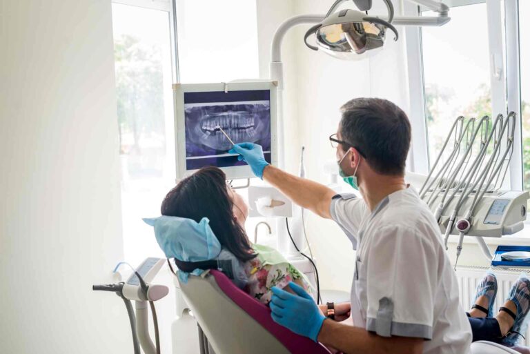
If you’ve recently had dental X-rays taken by your general dentist and are now being asked to take new ones at your endodontic specialist’s office, you might be wondering — why repeat the process? Aren’t all dental X-rays the same?
It’s a fair question, and one many patients ask. The short answer is: not all X-rays provide the same level of detail, and endodontic treatment requires a much closer look. Understanding the differences in dental imaging can help you feel more confident in the steps your specialist is taking to protect your oral health.
What Are Dental X-Rays, and Why Are They Used?
Dental X-rays (radiographs) are one of the most important tools in modern dentistry. They allow providers to:
- Bite misalignment — When the teeth don’t line up properly due to crowding, gaps, or jaw structure
- Detect cavities between teeth
- Evaluate bone levels and jaw structure
- Identify infections or abnormalities
- Track the development of teeth and roots
- Plan restorative or surgical treatment
General dentists typically use 2D digital X-rays, which are fast, non-invasive, and excellent for routine exams or cavity checks. These images offer a flat, two-dimensional view of your teeth and jaw.
For most dental visits, that’s all you need. But when the situation becomes more complex—such as when a root canal is being considered—more advanced imaging is often necessary.
Why Endodontists Need More Than 2D X-Rays
Endodontists specialize in diagnosing and treating problems deep within the tooth, particularly involving the roots and surrounding bone. While 2D X-rays are a helpful starting point, they often don’t reveal the full picture needed to make an accurate diagnosis.
For example, a 2D image may not show:
- Tiny cracks — Hairline fractures or microcracks invisible on standard X-rays
- Hidden canals — Some teeth have extra or oddly shaped root canals
- Infections in surrounding bone — May go undetected in flat images
- Root curvature — Important for treatment planning and avoiding complications
- Precise lesion depth — Helps determine the extent of infection or decay
In these situations, even a high-quality traditional X-ray can miss critical information.
What Is a CBCT Scan?
To overcome the limitations of 2D imaging, endodontists often use a CBCT scan, short for Cone Beam Computed Tomography. This type of imaging uses a cone-shaped X-ray beam to create a 3D image of your teeth, roots, bone, and surrounding structures.
Think of it as going from a photo to a 3D model — it’s a much more complete and detailed way to examine the inner workings of your mouth.
With CBCT technology, your endodontist can rotate and view the image from multiple angles, allowing for:
- Precise diagnosis — Better visibility of cracks and root abnormalities
- Early detection — Finds infections or bone loss before symptoms worsen
- Better treatment planning — Reduces surprises and complications during procedures
- Fewer surprises — A more predictable and comfortable patient experience
CBCT vs. Traditional X-Rays — What’s the Difference?
| Feature | Traditional X-rays | CBCT Scan |
|---|---|---|
| Image Type | 2D | 3D |
| Detail Level | Basic tooth structure | Detailed view of roots, bone, nerves |
| Best For | Routine dental checkups | Complex diagnoses and treatment planning |
| Limitations | Overlapping anatomy may hide issues | Reveals fine details from every angle |
While both types of images are important, they serve different purposes. Your general dentist uses traditional X-rays to keep up with overall oral health, while your endodontist uses CBCT to diagnose and treat specific issues with precision.
Is It Safe to Take a CBCT Scan?
Understandably, many patients are concerned about radiation exposure from repeated X-rays. The good news is that CBCT scans are designed to use a low dose of radiation, often comparable to or only slightly higher than standard dental X-rays.
Because CBCT imaging is so targeted, it can reduce the need for additional X-rays later in the process. Plus, having a clear and complete picture from the start means your endodontist can:
- Diagnose the issue more accurately
- Avoid unnecessary treatment
- Increase the chances of long-term success
Will My Dentist’s X-Rays Still Be Used?
Absolutely. Your general dentist’s X-rays are still an important part of your dental record. They help provide an overview of your oral health history and are often reviewed alongside the CBCT scan to ensure nothing is overlooked.
However, for a root canal or re-treatment, your endodontist needs that 3D view to ensure no cracks, infections, or hidden canals are missed. It’s not about repeating tests — it’s about refining the diagnosis.
Can’t My Dentist Just Do a CBCT?
CBCT technology is highly specialized and not typically found in general dental offices. Because of the cost and training required, it’s most commonly used in specialty practices — such as oral surgery, implantology, and endodontics.
When your dentist refers you to an endodontist, they’re ensuring that you have access to the best diagnostic tools available for the type of care you need. It’s a team effort — your general dentist starts the process, and your endodontist follows through with the deeper diagnostics and treatment.
Final Thoughts
Getting new X-rays at your endodontist’s office doesn’t mean your previous dental care was lacking — it means your specialist is using the most advanced tools available to make an informed, accurate diagnosis. 3D imaging like CBCT allows for a more complete understanding of your tooth’s condition, which leads to better results and a smoother treatment experience.
Need Expert Root Canal Care in Pensacola?
If you’re facing a possible root canal and want clarity about your diagnosis and treatment options, the team at Elite Endodontics in Pensacola is here to help. Our board-certified endodontists use advanced 3D imaging to ensure every detail is visible before starting treatment, so you can feel confident you’re in the best hands.
Contact us today to schedule your consultation — your comfort, clarity, and care come first.